Focused Ultrasound: A New Approach to Treating Cancer and Non-Healing Chronic Wounds
Wednesday, October 10, 2018
Five years ago, Dr. Ashish Ranjan established a focused ultrasound program at Oklahoma State University’s Center for Veterinary Health Sciences.
“Building on my prior work with high intensity focused ultrasound or HIFU technology at the National Institutes of Health, my laboratory initiated several funded research projects in cancer bearing rodent models to understand the feasibility of translating this approach to treat veterinary cancer patients,” explained Ranjan, BVSc, PhD, Kerr Foundation Endowed Chair and associate professor in the Department of Physiological Sciences. “Specifically, the projects aimed to tailor the HIFU sound energy for enhancing localized tumor killing, enhancing chemotherapy delivery, and optimizing the immune system for robust therapeutic outcomes. In addition, we worked on devising new methodologies for improving sensitivity of drug resistant pathogens to antimicrobials. Based on the promising data in rodents, the laboratory was recently funded by the Focused Ultrasound Foundation to conduct clinical trials in dogs with cancer and non-healing wound infections. That’s how we got started.”
Oklahoma State University is the first veterinary school to offer focused ultrasound treatment as a service in addition to surgery and chemotherapy.
“The HIFU service is currently available to owners for locoregional treatment of cancerous tumors, infected soft-tissue, and bone infections. Patients entering the clinical trial must meet the inclusion criteria of an active infection or the presence of a locally accessible tumor. The FDA is currently reviewing our application to include in the HIFU regimen nanoparticle immune adjuvants, which we developed in the lab. This will be especially beneficial for patients with aggressive cancer that has spread to other parts of the body from its primary site,” continued Ranjan. “While we are the only school to provide HIFU as a service, Virginia Tech also has a grant that is supporting research in focused ultrasound technology for veterinary cancer patients.”
Others working on this project at OSU include Drs. Danielle Dugat, Jerry Malayer, and Jerry Ritchey. Malayer provides cell and molecular biology support looking at the interactions of molecules in and on the cells that mediate the processes of tumor destruction while Ritchey is responsible for immunopathology support.
“I look at microscopic samples of the cancer to determine whether the cancerous tissue is being affected by Dr. Ranjan’s treatments to perhaps verify if the treatment is working or not,” said Ritchey. “We also run samples through a flow cytometer which gives us a picture into the function of the patient’s immune system during the cancer treatment because some of Dr. Ranjan’s therapies are aimed at enhancing the patient’s own immune system to help fight the cancer.”
“My role in the focused ultrasound clinic at the Hospital is to engage clients,” said Danielle Dugat, DVM, MS, DACVS (Small Animal), Cohn Family Chair for Small Animal Care, and assistant professor of small animal surgery in the Department of Veterinary Clinical Sciences at the veterinary center’s Veterinary Medical Hospital. “Dr. Ranjan and I seek out patients who are in need of this therapy. Together with others involved in the project, Drs. Kalyani Ektate, Harshini Ashar, and Donald Holter, we help manage these cases from the time they come in the door including their hospital stay, evaluations, taking measurements, and performing diagnostics—basically all the clinical aspects of maintaining that patient around the actual procedure itself.”
The HIFU procedure typically requires that the patient be very still for about an hour. Patients receive anesthesia and pain control during each procedure.
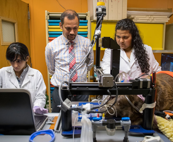
“Our state of the art system comes equipped with an imaging and treatment transducer. We use the ultrasound imaging transducer to locate the tumor,” stated Ranjan. “Once we have localized the tumor, we set boundaries for treatment, which we call a region of interest. Typically we have an image screen where we can see the cancerous mass and then we administer treatment to those regions of interest using the built-in therapeutic HIFU system. Depending on the disease profile, the energy from the therapeutic transducer is altered to attain various effects in the region of interest including local mild heating, tissue damage, and non-heat related cellular and bacterial stress. We call these kinds of therapy image guided therapy. We are doing the HIFU treatment under image guidance, which in this case, happens to be ultrasound.”
During this clinical trial phase, the veterinary center team reports that some cases have been successful and some have not.
“In some cases we had complete remission,” reported Ranjan. “The tumor was gone after one or two treatments. In other cases we had control of the disease. In other words, the tumor did not grow beyond what it was when the patient came to us, so that is also success.”
“My experience currently with the clinical trial cases has been very rewarding,” said Dugat. “We’re learning as we go what type of cancers may be more responsive or less responsive. Through this trial we are gaining information on how different tumors may react. Two cases that pop in the top of my mind are ones where the tumors have completely gone away. So for a patient where maybe surgery would have meant removing half of their jaw or reconstructing their lip, now they didn’t have to have any surgery and the tumor was removed via this method. That is the real rewarding part of this technology. When we know more information in the future, then maybe we can offer this as a first step or a first line of treatment before we even think about surgery.”
The two most critical benefits to focused ultrasound treatment over traditional treatments like surgery, chemotherapy, or radiation are that it is non-invasive and non-toxic.
“Focused ultrasound doesn’t require the use of scalpels and invasive surgeries to remove tumors or cure infections,” said Ranjan. “The other advantage would be it is non-toxic. It is safe to the patient so it can be non-invasively administered rapidly in a non-toxic manner.”
Cost-wise, there are some benefits to this treatment as well.
“As we are the first veterinary school to offer this kind of treatment, we also happen to be the first college to set the cost of the treatment,” explained Ranjan. “If an owner were going to go with focused ultrasound in contrast to surgery, they would be saving at least 50 percent of the treatment cost. A typical surgical procedure for an oral cancer would cost about $5,000, whereas in the case of focused ultrasound that would be available at $2,000 to $2,500.”
“This technology is very exciting,” said Dr. Jeff Studer, Hospital director. “It, combined with the tireless efforts of Drs. Ranjan and Dugat, is providing treatment options for our patients who would have otherwise not had options.”
According to Drs. Dugat and Ranjan, owners have been very willing to participate in the clinical trial.
“Owners are happy to try to advance medical care not only to get good results in their own patient but to give us more information so we can help future patients,” said Dugat. “So it’s been very positive even in the cases that haven’t worked. The owners have been very thankful that they have gotten the chance to try and see if there was anything that could be done.”
“It was pretty fabulous that Laddie was part of that trial, mostly financially so he could be treated,” said Jennifer Reyna of Stillwater, Okla. “He is doing just fine now.”
Laddie is a 10-year-old Border Collie who had a mass on his mandible. As part of the cancer clinical trial, treatment costs were covered by OSU endowed chairs. The mass was confirmed to be an acanthomatous ameloblastoma. Laddie received one focused ultrasound treatment for three to five minutes non-invasively. Within a few days, the mass fell off. A recheck three weeks later showed Laddie was cancer free with no ulcers present. No evidence of neoplasia or ameloblastoma were found in the diagnostic evaluation.
Oreo is another success story. Lance Millis, director of student academic services at OSU’s College of Engineering, Architecture and Technology, has been bringing Oreo to OSU’s Veterinary Medical Hospital since the 9-year-old Shetland Sheepdog was a puppy. When his veterinarian, Dr. Paul DeMars, noticed a mass on Oreo’s lower right lip, Oreo was referred to the Hospital’s dentist who surgically removed the mass. A biopsy showed it to be plasmacytoma. When the mass returned, Millis had the opportunity to enter Oreo in the clinical trial.
“Oreo is doing great,” said Millis. “His demeanor has been terrific. There has been no recurrence; he’s doing awesome. When we would take Oreo in for his treatments, the vet students would recognize him. Dr. Dugat would ask about him. We’re very happy he has friends at the Hospital.”
Oreo received two focused ultrasound treatments for three to five minutes each non-invasively. A recheck three weeks later showed no cancer with no ulcers present.
“We have now expanded the treatment from dogs to cats,” reported Ranjan. “There is a significant amount of interest at the Hospital to do horses as they also get skin cancer or sarcoids. My lab is currently working on developing a system for that kind of treatment. These translational projects meet the OSU mission of developing clinically relevant technologies that enhance non-invasive and minimally invasive treatments. The owners currently see a lot of benefit in having a treatment like this where there is no surgery, no infection. It has very minor complications, and it’s relatively rapid.”
“The future appears bright when it comes to the possibilities,” echoed Dugat. “We want owners to know that there are newer methods out there whether it be for chronic wound healing or treating cancer.”
“As we learn more about this approach, we are optimistic at the possibilities that this technology can offer. I give credit to several undergraduate, graduate, and DVM students who worked tirelessly to bring this to fruition. It’s said that the best reward in biomedical sciences is when research is translated from bench to bedside. Our early data in the canine patients represents such a vision, however, a lot of research is still needed to be done to establish the true feasibility of this technology, and wide-range clinical use for a variety of indications,” added Ranjan.
If you would like more information on OSU’s focused ultrasound program, please contact Dr. Ranjan at ashish.ranjan@okstate.edu. If you would like to support veterinary medical research, please contact Ms. Chris Sitz, senior director of development and team lead for the veterinary center with the OSU Foundation, at 405-385-5170 or csitz@osugiving.com.