
Handling canine MEC
Monday, October 7, 2019
Some dogs are born with abnormal protrusions on their heads; others sustain them after trauma or surgery or as a result of high blood pressure in the brain. And these protrusions can be harmful.
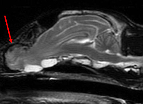
These protrusions that a dog is born with occurred as its central nervous system developed and its skull became somehow malformed. These intracranial malformations can cause canine ethmoidal meningoencephaloceles (MEC). The intracranial congenital malformation results from an incomplete closure of the calvaria, which causes a protrusion of fluid, brain tissue and meninges (MEC) or of the meninges only — in other words, a meningocele (MC). MECs may also occur after trauma, surgery or as a result of chronic intracranial hypertension.
Burmese cats have shown a hereditary predisposition to MECs, but nutritional deficiencies and teratogenic factors may play a role in the development of the malformations.
Few cases of MEC exist in veterinary literature.
If the congenital protrusion of the brain tissue is covered by skin, the abnormalities are most often detectable from birth as a fluctuant swelling along the top of the head. If the MEC is encased within the boney nasal or ethmoidal regions, the protrusion may only be detected with cross-sectional imaging such as CTs or MRIs.
Dogs with MEC typically exhibit epileptic seizures and/or abnormal behavior. Seizures are the most common signs in young animals (up to 6 years old). Additional signs may include aggressiveness, compulsive behavior, hyperactivity, intermittent yelping, star-gazing or fly-catching.
Reported cases have shown a herniation of the olfactory bulb through a cribriform plate defect. The cribriform plate is a sieve-like bone that allows passage of the nasal receptors to cross into the olfactory bulb for interpretation of smell. With the protrusion of the olfactory bulb, additional brain material may be stretched or protruded out of the cranial vault. The protruded part often acts as a breeding place for seizure activity.
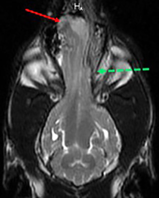
The most frequent location of the MEC is in the nasal cavity, with the ethmoidal bone affected the most, followed by the parietal bone and the frontal bone. The size of the protrusion and the contents play a role in the decision for medical or surgical management. Mild herniation may be well-managed with medical options, whereas moderate and severe herniation may be better treated with surgery.
This is commonly addressed in treating people with MEC.
Both medical and surgical corrections have been implemented in canines.
Medical management of mild MEC has shown promising results, including reducing seizures or showing more normal behaviors as the main goal. The greatest assessment risk of medical management is increased infection leading to meningitis.
Surgical management of moderate to severe MEC involves excising the protruded portion of the brain, reconstructing the cranial vault and creating a watertight closure of the dural defect. The surgery aims to reduce seizures and prevent a progression to meningitis. The surgical treatment of MEC is the same for people.
STORY BY: Carrie Kuzma, DVM, an assistant professor of radiology in the diagnostic imaging section at OSU’s Veterinary Medicinal Hospital. She is eligible to sit for the board examination to become a diplomate of the American College of Veterinary Radiology.