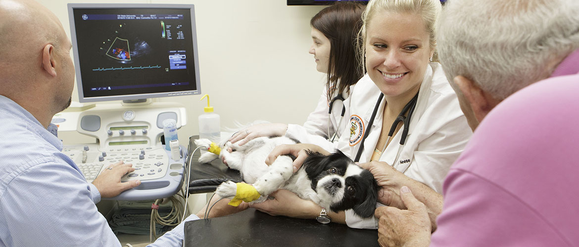
Ultrasound offers versatility in veterinary medicine
Monday, July 1, 2019
Ultrasound, also known as sonography, has been used in human and veterinary medicine alike for many years. Most people are familiar with its use in prenatal exams, allowing a “sneak peek” of the baby in utero. Ultrasound has a multitude of other uses and is becoming increasingly available to veterinary patients throughout Oklahoma.
An ultrasound exam is performed by using a computer with specialized software with an attached “probe” or transducer. The surface of the transducer is placed on the skin (often clipped in veterinary patients) with a topical gel to eliminate any air between the skin and the probe surface. Tiny vibrations within the probe send ultrasonic sound waves into the tissue. These waves then “bounce” back to the probe, which use that information to produce an image. This process is painless, involves no risk and can be performed quickly by a trained professional. Patients are often examined while awake or may be sedated to help them lie still.
Ultrasound is used in veterinary medicine in a variety of ways. In companion animals such as cats and dogs, the most common use is examining the abdomen in patients that present with GI signs such as vomiting or lack of appetite, urinary signs such as straining or increased amounts of urination, or elevations in blood work values that indicate liver or kidney disease. Ultrasound is sensitive to changes in small organs such as the adrenal glands, the gall bladder and the pancreas, which cannot be assessed with other routine procedures such as X-rays.
Ultrasound’s versatility allows it to be used in a wide variety of cases in veterinary medicine. For example, ultrasound is used to examine horses that present with signs of colic. Lame or limping animals, such as working horses, can have an ultrasound evaluation of the tendons and ligaments of the limbs, which can easily detect swelling or tears. Ultrasound examination of the heart, called echocardiography, can detect the causes of heart murmurs, arrhythmias and other heart diseases. It can also look into the thorax of animals that have fluid accumulations around the lungs.
The major limitation of ultrasound is that these very-high frequency sound waves cannot travel through air so only the surface of the lungs can be evaluated, and X-rays are still used routinely to evaluate the thorax. Likewise, abdominal examinations in large patients with large amounts of gas in their stomach and intestines may be limited. Another important limitation of ultrasound is that it is very dependent on the skill and experience of the operator, and ultrasound examinations can vary widely between individual users.
Ultrasound can be used on any species, from the smallest birds to the largest rodeo bulls. Essentially, anywhere you can get the transducer onto the skin of a patient, you can perform an ultrasound exam. Ultrasound is even used to look through the eyes of patients with cataracts, in order to make sure the retina is healthy before performing cataract surgery. Ultrasound can accurately guide needles into tissues such as tumors to obtain a sample for diagnosis.
Ultrasound equipment is becoming more available to veterinarians, and many general practice veterinarians have the equipment in their practices. Veterinary students are offered elective courses in sonography, and continuing education programs throughout the country offer practicing veterinarians hands-on training. Ultrasound is generally available in all specialty and emergency veterinary hospitals, where in-depth examinations may be performed by specialists such as veterinary internists, criticalists, and veterinary radiologists.
In short, ultrasound is a powerful tool available to the veterinarian and pet owner that can provide important information about the inner workings of your animal in order to provide advanced diagnosis and improved health care to animals of every shape and size.
