Professor named Fellows Grant Award recipient
Thursday, February 27, 2020
Dr. Paul Gignac, associate professor in Anatomy and Cell Biology at OSU Center for Health Sciences, is the recent recipient of a 2020 Fellows Grant Award from the American Association for Anatomy.
Gignac is one of only two recipients of the $25,000 grant this year, which will be used to further his research of diceCT imaging and its applications in the field of neuroscience.
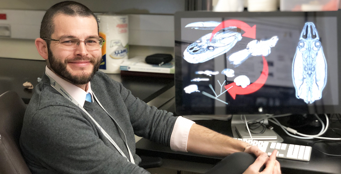
DiceCT— diffusible iodine-based contrast-enhanced computed tomography imaging— is an ex-vivo process that allows researchers and scientists to use X-ray technology to create intricate 3D images of soft tissues in all animals, Gignac said.
“Usually you can’t see soft tissue very well in X-rays, but with diceCT, you get almost everything. You can see aspects of brains, muscles, organs. They’re all digital and spatially accurate,” he said. “This allows us to more rapidly see and study whole brains, from hagfishes to humans, at a scope that we’ve never had before.”
In the past, the only way to study brain tissue at this scale was to physically dissect brain slices, manually align them, and then look across each section one and a time.
“It would take several months of work to produce these brains sections, but we can do this in a matter of days to weeks,” Gignac said. “The biggest advantage of micro-CT imaging is that we can produce the highest resolution whole-brain images of any technology, which translates to capturing finer gray- and white-matter detail and all of the organ.”
This can be helpful when understanding the effects of brain trauma in cadaveric samples, for example.
"We can look, not only at the location of the primary trauma, but elsewhere in the brain where there may be co-factors, or where we can identify other areas that might also be injured or affected."
There’s been a lot of interest in the diceCT technique, Gignac said, but the biggest skeptics seem to come from the area of neuroscience, which is where he hopes utilizing the Fellows Grant can help bridge that gap between morphology and neurology.
“I have a vision of using diceCT in conjunction with all the tools neuroscience already has,” he said. “This tool is inexpensive and relatively rapid. It could be a kind of digital ‘hub’ that connects all the others.”
Gignac said the goal is for the research he and his team are conducting and their findings to be submitted for a 2021 National Science Foundation grant.
“This kind of project is an excellent example of how anatomy research can unite different wings of the biomedical sciences,” he said.
MEDIA CONTACT: Sara Plummer | Communications Coordinator | 918-561-1282 | sara.plummer@okstate.edu