Inside Your Animals
Wednesday, September 28, 2016
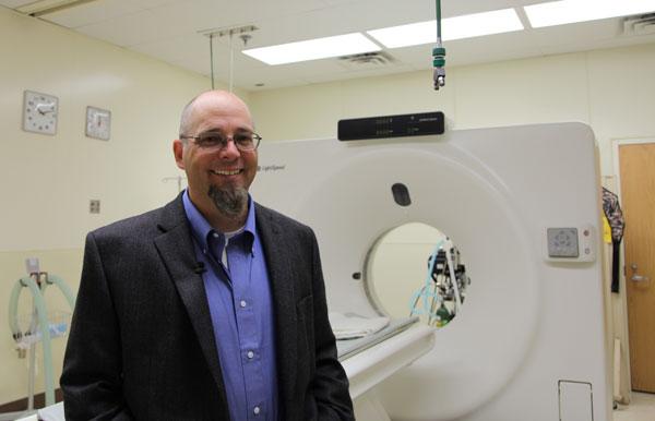
Dr. Corey Wall is a veterinary radiologist at Oklahoma State University’s Center for Veterinary Health Sciences. He leads the digital imaging services provided by the center’s Veterinary Medical Hospital.
“We have several pieces of imaging equipment here at Oklahoma State University,” said Wall. “We have three radiology suites where we take x-rays of our veterinary patients. One is an equine specific suite. We also have two small animal suites. In addition, we have a mobile radiographic capability where we can go stall-side or potentially out to a trailer.
“The hospital is equipped with a CT scanner,” he continued. “It’s a 4-slice GE helical scanner. We have a 1.5 Telsa MR unit. We also have a nuclear medicine laboratory where we do equine bone scans and feline thyroid studies and treatments. Additionally, we do a large amount of small animal and exotic abdominal ultrasound imaging. We also have a fluoroscopic unit where we can take moving x-rays of a small animal patient.”
Wall’s team acts as an ancillary service to the primary veterinary clinicians. There are two radiologists and three technicians. The newest member of the team is Dr. MacKenzie Hallman. She just completed her radiology residency at Kansas State University.
“In small animals we can image any area of concern or organ system that the primary clinician is worried about with their patients. Mostly what we are looking at is an anatomic change – somehow the shape or structure of that organ has been altered. Also with the nuclear medicine laboratory we can do some physiologic imaging of various organ systems, this allows us to get a better understanding of how well the organ of concern is functioning in the patient,” he added.
According to Wall, the most common procedure performed is thoracic radiography.
“Taking a chest x-ray of dogs and cats that have coughing episodes, heart disease or cancer that may have spread throughout the body seems to be our most commonly requested examinations,” stated Wall. “The most unusual case that I have seen recently on an MR was a cat with multifocal brain and meningeal disease that also has some fairly extensive disease within its sinuses and nasal cavities.
“The greatest thing about digital imaging is that the information gets disseminated everywhere,” he said. “So it makes consultations much more beneficial to the pets. In the past, when we had film, there was only one set of images and that film had to go everywhere and be tracked down. That can be lost, misplaced, or squirreled away for some sort of teaching purposes and is essentially hidden from all other viewers. With digital radiography, it goes to a server and anybody with a computer or device has the potential to bring up that image.
“For example, an image is disseminated throughout the hospital system so multiple clinicians can be looking at it. The surgeon can be looking at it in the surgical suite, while students are viewing the study in the rounds room. So I think the educational aspect and the dissemination of that information makes digital imaging such a huge advancement for our profession.”
And that change over the last few years to digital technology has benefitted the general practitioner as well.
“The biggest thing that digital imaging has changed is the ease of consultations from the general practitioner’s standpoint,” explained Wall. “In the past, with film they would have to be FedExed to a radiologist to be viewed, interpreted and usually followed up with a phone call to try to get that information returned to the practitioner as quickly as possible for that pet. Now they can be sent through the internet, pulled up at a work station and a phone call or an email sent off in several minutes. So the turnaround time is now hours versus days.”
As wonderful as digital imaging is, there is a shortage of radiologists in the field.
“There is a huge shortage of radiologists in the field of veterinary medicine at this point in time,” said Wall. “It’s multifactorial but it is an issue that the American College of Veterinary Radiology is working on improving. I feel real fortunate to be a radiologist and a diagnostic imaging specialist. I am one of those lucky individuals who enjoys what they do every day that they do it.”
Wall earned his DVM degree from Colorado State University (1999). In 2010, he earned a master’s degree and completed his residency in diagnostic imaging at the University of Missouri and became a diplomate of the American College of Veterinary Radiology. He joined the faculty at OSU in 2014.
For more information on the diagnostic imaging services available at OSU’s Center for Veterinary Health Sciences, visit https://cvhs.okstate.edu/osu-veterinary-medical-hospital/diagnostic-imaging.