OSU Veterinary Medical Teaching Hospital saves bucking bull’s jaw
Thursday, October 24, 2024
Media Contact: Taylor Bacon | Public Relations and Marketing Coordinator | 405-744-6728 | taylor.bacon@okstate.edu
Nothin’ But Try Ranch, owned by Chad and Jenny Drury, is in Erick, Oklahoma, and is home to multiple world-renowned bucking bulls.
When Mouse Trap, a 3-year-old bucking bull, didn’t come up to eat one night, the Drurys knew something wasn’t right. After finding Mouse Trap with excessive salivation and noticing he could not eat, the Drurys called their local veterinarian.
When they examined swelling on the right jaw, radiographs showed Mouse Trap had fractured it. Their local veterinarian referred Mouse Trap to the Oklahoma State University Veterinary Medical Teaching Hospital for surgical repair.
After arriving at OSU the next day, Mouse Trap was given a complete physical exam, and further radiographs were taken to ensure the ideal plan for surgical repair.
“The fracture on the right was complete with a second smaller fracture next to it,” said Dr. Andrea Messina, veterinary clinical sciences intern. “We were concerned about possible tooth root involvement as the main fracture line appeared to involve the roots of the third premolar.”
Unilateral, or one-sided, jaw fractures don’t always require surgical repair. However, Mouse Trap’s unwillingness to eat warranted the surgery. Surgical repair would help stabilize the jaw during the healing process.
“The surgical repair plan consisted of an external fixation device, which involves the use of metal pins drilled into the bone, which are connected by a rod that acts as a scaffold. An oral cerclage wire was also planned for additional stabilization,” Messina said.
Once in surgery, Messina and her team placed this wire around the first through third premolars on the right side of the jaw. After the wire was placed and the jaw was stable, the ends of the wires were cut and covered with Technovit, a resin, to prevent the wire from damaging the inside of the mouth.
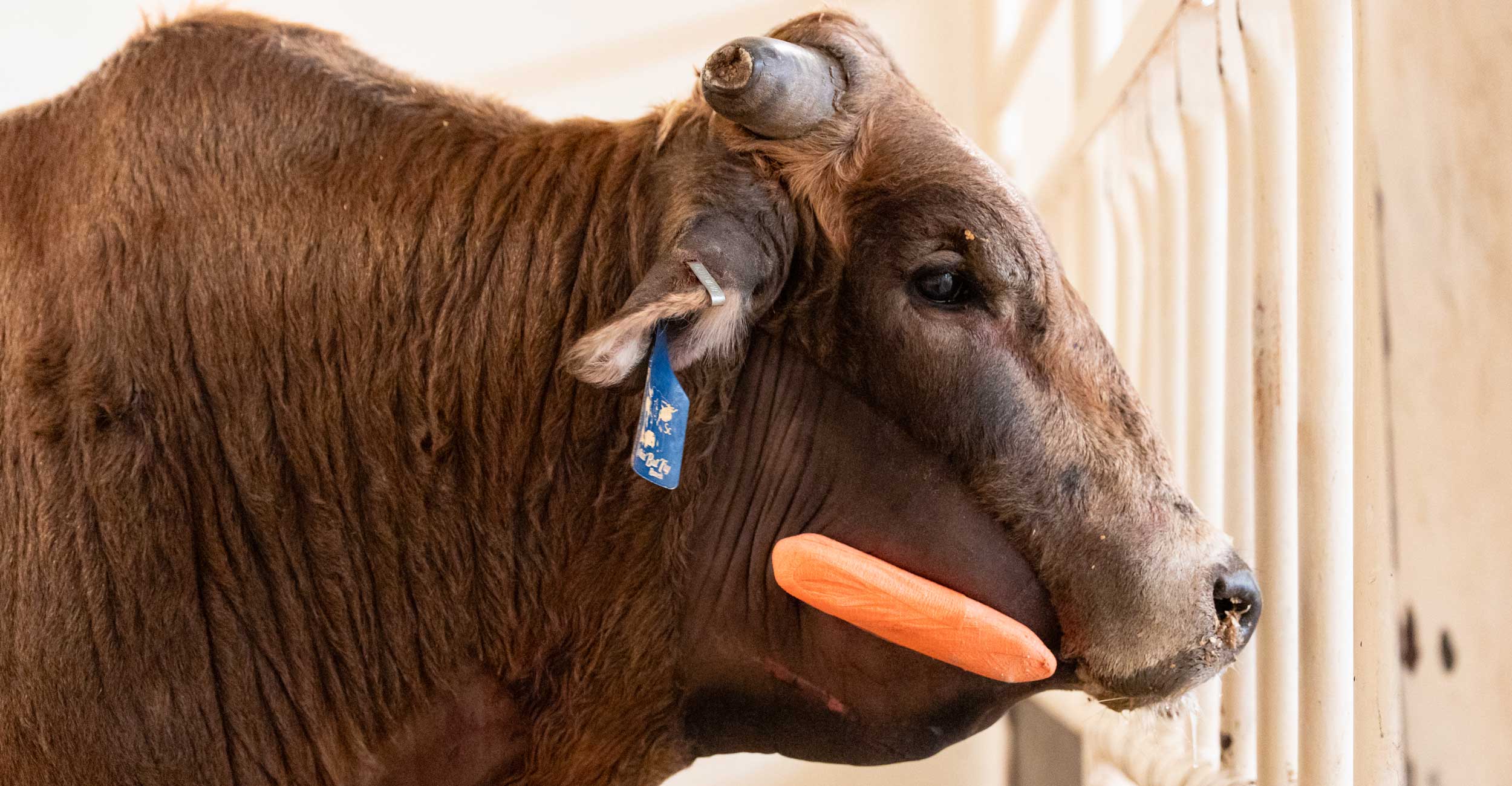
Messina and her team used intra-op radiographs to place hypodermic needles along the jaw to confirm pin placement before making the incisions for the pin’s sites. After four pins were placed, a carbon fiber connecting rod was placed to stabilize them. After placing the pins and the rod, it was bandaged with gauze and orange vet wrap.
“What blows my mind is that Mouse Trap went from not being able to eat at all to eating as soon as he woke from surgery,” Drury said.
Less than 24 hours after surgery, Mouse Trap returned to eating, drinking and tolerating his pins well. After three days, Mouse Trap was discharged to go home with a recheck appointment scheduled for six weeks out. At his six-week post-op appointment, it was confirmed that the fracture site had thickened and was firm because of new bone growth.
As a result of his quick, six-week recovery, Mouse Trap’s pins were removed during his recheck appointment.
“Everything went great, and he is completely healed,” Drury said. “We plan to start exercising and getting him back into shape so he can start bucking again.”
Photos By: Taylor Bacon
Story By: Kinsey Reed | Vet Voices Magazine
