Taking the Guesswork out of Equine Lameness Detection
Monday, January 4, 2021
Lameness is the most common ailment that horses suffer. In fact, lameness is estimated to cost U.S. horse owners more than $1 billion every year.

Lameness can be described as an abnormal walk, usually noted by an asymmetry in movement. However, lameness is not a disease; it is a clinical sign resulting from pain or dysfunction of the locomotor system. The source of lameness must be localized to a specific area for an accurate diagnosis. Often, localizing the source of lameness can be challenging.
To identify it, a veterinarian commonly uses the horse’s age, breed and discipline as well as its historical information in combination with clinical examination findings. The lame limb and the severity of lameness is determined by the veterinarian using a 0-5 grading scale from the American Association of Equine Practitioners — 0 is not lame and 5 is non-weight-bearing lame.
Veterinarians develop a keen eye allowing them to recognize the mildest of lameness. However, research has shown that there can be much discrepancy between veterinarians as to the severity and even sometimes the limb displaying the lameness when evaluating the same horse. Fortunately, there are technologies that can provide objective information to assist in lameness detection.
One of these technologies is a force plate, an instrument that measures the amount of force applied to it. In sports medicine, this force is termed the ground reaction force. The force plate has long been considered the gold standard for equine lameness detection. Lameness is often inconsistent, so multiple measurements must be collected to determine the average ground reactive force of each limb.

Generally, the average ground reaction force exhibited by each limb should be similar from side to side. If there is a significant difference, the limb exhibiting the lower ground reactive force is designated the lame limb. Although subjective evaluation and the force plate are both valid methods of lameness assessment, there is little correlation between the grading scale and ground reaction force measurements. This is because these methods are evaluating different movement parameters. The force plate measures the force of movement, otherwise known as kinetics, where the human eye evaluates the geometry of movement, or kinematics. Due to the time and effort required to obtain the required measurements, the force plate is not often used in clinical lameness evaluations, but it is a valuable research tool.
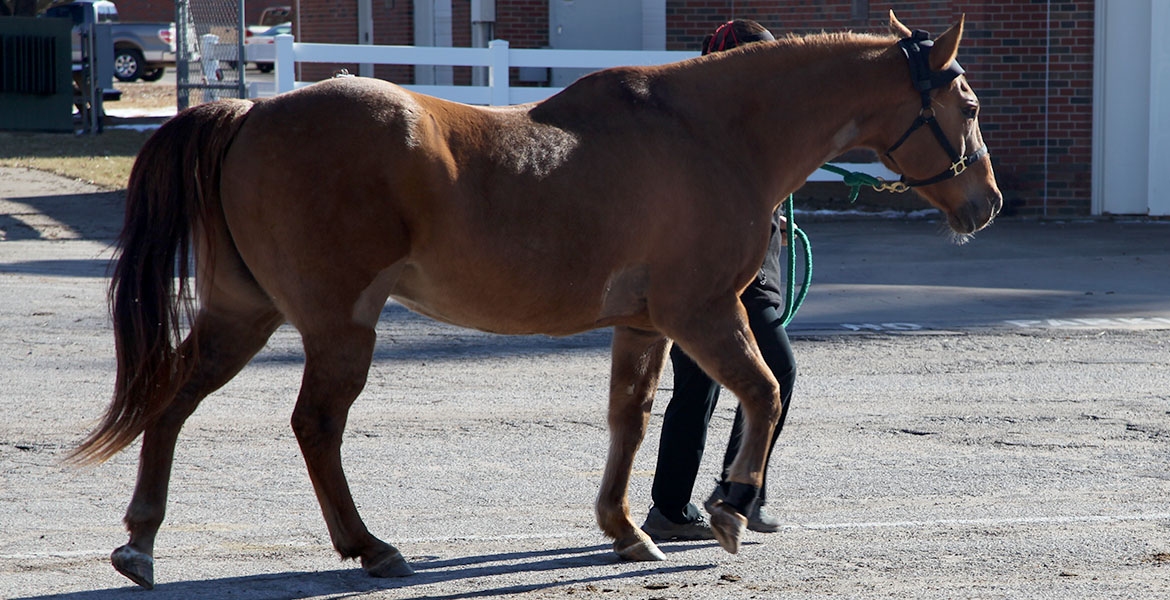
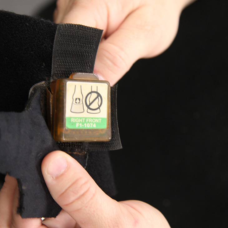
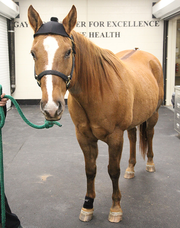
Inertial sensors offer another objective way to evaluate lameness in horses. They can measure movement down to a fraction of a millimeter at 200 times per second, far more sensitive than the human eye. Once attached to specific locations, inertial sensors record the movement patterns of a horse over 25 to 35 strides and communicate this information to a computer program, which then generates a report describing any asymmetries. Veterinarians can use this information to help determine if a horse is lame, which limb is lame and the severity of the lameness. Inertial sensors can also be used to evaluate response to flexion tests, improvement following local anesthesia or to document treatment success at re-evaluations that may be weeks to months later.
Inertial sensors have been validated by several research studies using both subjective assessments and force plate analysis. This methodology more closely correlates to subjective analysis because both evaluate for lameness in a similar fashion, through kinematics. Inertial sensors are easily utilized in a clinical lameness examination because the sensors can collect data simultaneously with the veterinarian’s subjective evaluation. This information, combined with the clinical exam, can provide a more complete assessment of a horse’s lameness.
The Equine Surgery and Sports Medicine Service at the OSU Veterinary Medical Hospital utilizes an inertial sensor system in addition to subjective evaluation for almost all lameness evaluations. Additionally, the OSU Equine Research Park is home to a force plate commonly used for lameness research. Not only do these technologies provide a better service to our clients and patients, but our veterinary students also benefit from comparative lameness analysis. They no longer have to rely and learn solely on the subjective opinion of a clinician. These technologies are not meant to replace the veterinarian’s role in the lameness evaluation but they can expose any errors or subconscious bias that may appear due to the subjective nature of the traditional lameness examination.
STORY BY: Mike J. Schoonover, DVM, MS, DACVS-LA, DACVSMR, an associate professor of equine surgery and sports medicine at the Oklahoma State University College of Veterinary Medicine. He is board certified in both equine surgery and sports medicine and rehabilitation.
