Veterinary Viewpoints: How deep puncture wounds in horses’ feet can be found, treated
Monday, November 15, 2021
Horse’s hooves are formed of insensitive, cornified material called horn and can be very resilient to penetration by an object. Inside the hoof capsule lies bone, joints, tendons, ligaments and other soft tissues.
While the hoof capsule is designed to protect these structures and provide the horse with a platform to support its weight, traumatic events can puncture the hoof and affect the deeper sensitive tissues. Puncture wounds often occur from stepping on nails, screws, or other such objects.
Sustaining deep puncture wounds to the foot causes lameness in horses. The opening of the puncture wound may or may not be evident during physical examination. The veterinarian examining the foot must try to determine if the horse is experiencing a common foot abscess or a puncture wound.
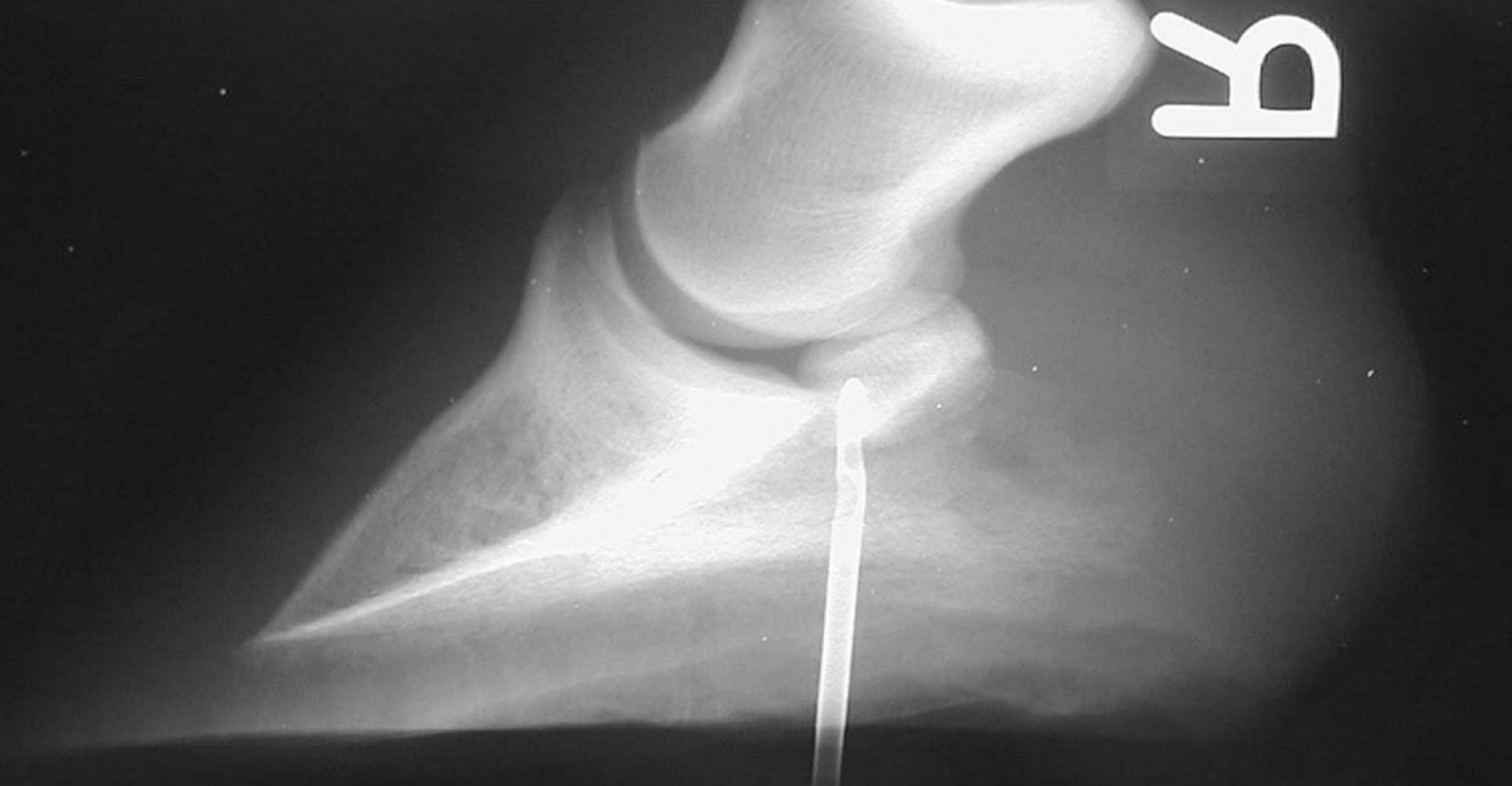
If a puncture wound is suspected, radiographs may be used to determine the location and depth of the injury. The veterinarian will place a metal probe into the wound tract and take radiographs of the foot. By doing this the veterinarian can better determine the location and depth of the puncture, as the metal probe will be seen as a bright white object on the radiographs.
To treat deep hoof puncture wounds, remove the foreign object, if it is still present, and debris from the puncture wound. This may involve surgery to clean and debride the wound. Horses who have sustained a deep puncture wound of the foot are treated with antibiotics to fight the likelihood of infection.
A tetanus toxoid vaccine is also given as early as possible after the injury occurs. Deep punctures create the ideal environment for the tetanus organism to grow and release its deadly toxin. Anti-inflammatory drugs, such as phenylbutazone, are also administered to relieve inflammation and pain.
During the healing time, the foot is wrapped in a clean, dry medicated bandage. If extensive removal (debridement) of the sole and deep tissues of the hoof was necessary to treat the puncture wound, a special shoe with a protective plate on the bottom called a medicine plate shoe is placed on the foot to protect the wound.
The plate is made so that bolts can be unscrewed to remove the plate from the shoe to medicate the wound. This treatment is continued until the wound is totally healed. Treatment could take several weeks. Prognosis for soundness is dependent on what underlying structures were involved and if infection was controlled.
Key take home message: If your horse suddenly becomes lame, a deep puncture wound to the hoof should be considered. If left untreated, puncture wounds to the foot may be disastrous. Call your veterinarian early. Prompt diagnosis and treatment may lead to a better outcome.
About the author: Daniel J. Burba, DVM, Dipl. ACVS, is a professor of equine surgery at Oklahoma State University’s College of Veterinary Medicine. He is a diplomate of the American College of Veterinary Surgeons and head of the Department of Veterinary Clinical Sciences at the college’s Veterinary Medical Hospital.
Veterinary Viewpoints is provided by the faculty of the OSU Veterinary Medical Teaching Hospital. Certified by the American Animal Hospital Association, the hospital is open to the public providing routine and specialized care for all species and 24-hour emergency care 365 days a year. Call 405-744-7000 for an appointment or more information.
OSU’s College of Veterinary Medicine is one of 32 accredited veterinary colleges in the United States and the only veterinary college in Oklahoma. The college’s Boren Veterinary Medical Hospital is open to the public and provides routine and specialized care for small and large animals. The hospital offers 24-hour emergency care and is certified by the American Animal Hospital Association. For more information, visit https://vetmed.okstate.edu or call 405-744-7000.
